– Ship Everywhere in Pakistan GraceSurgical.pk We want you to love our products. If you are not completely satisfied with your purchase you can return it free within 7 days, no questions asked. Learn more
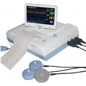
An ECG (Electrocardiogram) machine records the heart’s electrical signals via skin electrodes, translating them into a graph to check rhythm, rate, and function, revealing heart conditions. It works by picking up tiny electrical impulses from the heart, amplifying them, and displaying them as waves on paper or a screen, showing upward deflections for activity towards a lead and downward for activity away, helping diagnose issues like irregular beats or blockages. Standard 12-lead ECGs use 10 electrodes on limbs and chest for a comprehensive view.
How it works
Signal Collection: Electrodes (sticky patches) placed on the chest, arms, and legs detect the heart’s electrical impulses.
Amplification & Processing: Wires connect electrodes to the machine, which amplifies these small signals.
Recording: A processor converts the electrical data into a visual trace (waveform) on a screen or printed on graph paper.
Interpretation: Doctors analyze the waves (P, QRS, T) to assess heart health, looking for patterns indicating issues.
Key components & types
Electrodes: Sensors attached to the skin (10 for 12-lead ECG).
Leads: Different “views” (12 in a standard ECG) created by electrode combinations, capturing electrical flow from various angles.
Types: Beyond standard resting ECGs, there are Stress ECGs (treadmill/bike) and Holter Monitors (portable, 24-hour recording).
What it detects
Irregular heartbeats (arrhythmias).
Heart attacks or damage.
Problems with blood supply (ischemia).
Structural heart issues or electrolyte imbalances.


Reviews
There are no reviews yet.